
Ultra Structure of Animal Cells Microslide Manufacturer,Supplier and Exporter in India

Product Code : EL-CE-6749
Jlab Export is a leading Ultra Structure of Animal Cells Microslide Manufacturer,and suppliers in India
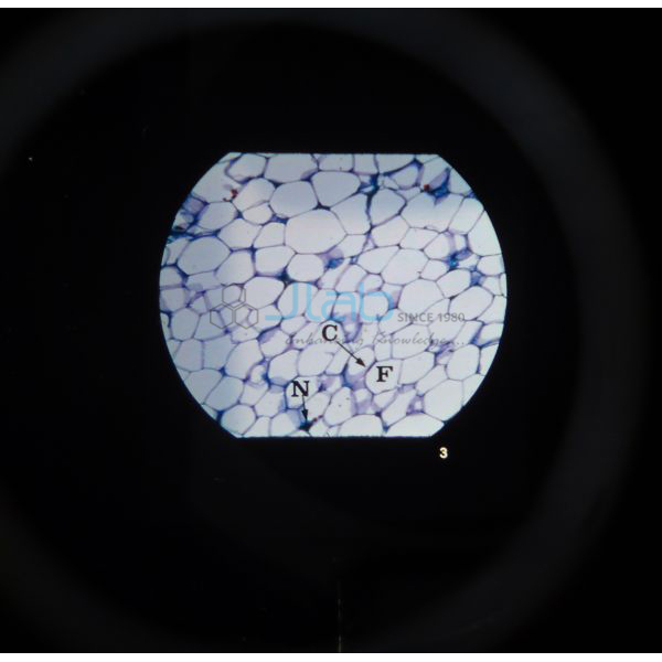
Ultra Structure of Animal Cells Microslide (code EL-CE-6749) is a Microslide set of eight related 35 mm images, photographed through a microscope, that shows the fine structure of animal cells — from the Golgi body and mitochondrion to the centriole, lamp-brush chromosomes and muscle cell fibrils. Arrows and callouts mark what students should find, and a text folder with a pocket comes with the set. JLab (Jain Laboratory Instruments Pvt Ltd, Ambala, India) supplies it to schools, colleges and dealers.
Specifications
| Parameter | Detail |
|---|---|
| Brand | Microslide |
| Format | 8 related 35 mm images photographed through a microscope |
| Images | Golgi body and vacuole; gland cells; plasma cell; mitochondrion; centriole; lamp-brush chromosomes; nerve cell membrane; muscle cell fibrils |
| Arrows and callouts | Help students locate the important features being studied |
| Text folder | Written to stimulate, inform and question students; has a pocket for storing the Microslides |
| Viewer | Viewed through a Microslide Viewer (separate item) |
| Quality check | Each set is visually inspected before dispatch |
| Pack quantity | Each (one set) |
| Minimum order quantity | 10 units |
| Dispatch time | Usually 7–10 working days |
| Product code | EL-CE-6749 |
| Supplied by | Jain Laboratory Instruments Pvt Ltd (JLab), Ambala, India |
What Does the Animal Cell Ultrastructure Set Teach?
“Ultrastructure” is the fine detail inside a cell, at the level usually studied with the electron microscope, and each of the eight images links a structure to its job. The Golgi body and the gland cells show how cells package and release secretions; the plasma cell is the white blood cell that releases antibodies; the mitochondrion, with its folded inner membrane, is where aerobic respiration releases energy; and the centriole helps organise the spindle when an animal cell divides. Lamp-brush chromosomes, the large looped chromosomes found in amphibian egg cells during meiosis, show genes being read, while the nerve cell membrane and the muscle fibrils, built from the proteins actin and myosin, connect the set to nerve impulses and contraction.
The topics follow the class 11 chapter on the cell and carry on into class 12 and college cell biology. A teacher can give each group a set and a viewer, ask students to name each organelle from the arrows and callouts, and then use the questions in the text folder for discussion or revision. Keep the Microslides in the folder pocket when they are not in use.
What to Specify When Ordering
- Quantity — the minimum order is 10 units, one set per group.
- Viewers — Microslide Viewers are quoted separately; state how many you need.
- Companion sets — the cell nucleus and cell structure Microslides cover the same unit at a simpler level.
Frequently Asked Questions
What organelles and cells does the ultrastructure Microslide show?
The Golgi body and vacuole, gland cells, a plasma cell, a mitochondrion, a centriole, lamp-brush chromosomes, a nerve cell membrane and muscle cell fibrils — eight images in all.
Do I need a special viewer?
Yes, the images are meant to be viewed through a Microslide Viewer. It is not part of this listing; JLab lists the viewer separately and can add it to your quotation.
Which level is it suited to?
Senior secondary and college biology: the organelles match the class 11 cell chapter, and topics such as lamp-brush chromosomes and muscle fibrils extend into college cell biology.
Who supplies the Ultra Structure of Animal Cells Microslide?
JLab — Jain Laboratory Instruments Pvt Ltd, Ambala, India — supplies this Microslide set and quotes for it directly.
Supplier
JLab is the trading name of Jain Laboratory Instruments Pvt Ltd, incorporated in 2017 and located on Hargolal Road, Ambala, Haryana. The company produces school and college lab apparatus and, to round out its biology range, also resells teaching resources of other brands such as these Microslide sets; every set is checked visually before it is dispatched. JLab exports worldwide and ships to buyers in every region — Ecuador in South America, Malaysia in Southeast Asia, New Zealand in Oceania, Nigeria in West Africa and Eswatini in Southern Africa are a few examples.
Related Cell Biology Resources
- Microslide Viewer — designed for viewing Microslides; a white reflector gives the light, so no batteries are needed.
- Microslide the Cell Nucleus — eight images on the nucleus, the nuclear membrane and the endoplasmic reticulum.
- The Cell Structure Microslide — whole cells such as cheek cells, cork, onion skin and bacteria.
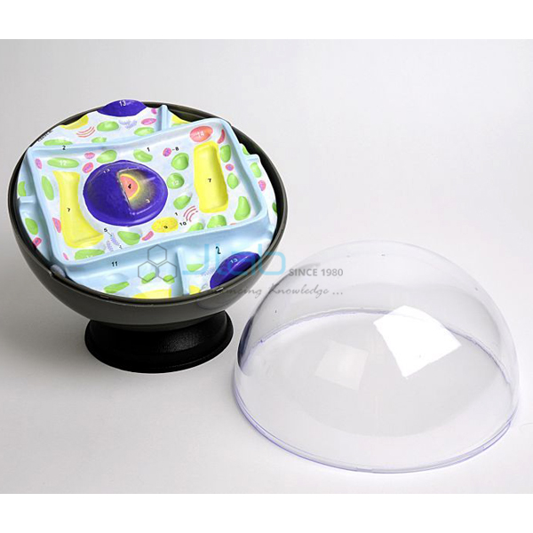
- Animal Cell Model — a model enlarged 20,000 times with mitochondria, Golgi apparatus, centrioles and lysosomes.
- Inflatable Animal Cell Model — a classroom visual aid for reviewing cell parts and building vocabulary.
More items are in Cell Equipments and the wider Biology Lab Equipment range.
Request a Quote
Quotes come directly from JLab. Click Add To Cart on this page, or use the contact page to send the number of sets (minimum 10 units), the viewers you need and your delivery location.
Last updated: 4 October 2026