

Microslide the Cell Nucleus Manufacturer,Supplier and Exporter in India
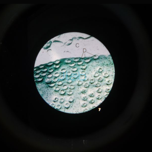
Product Code : EL-CE-6748
Jlab Export is a leading Microslide the Cell Nucleus Manufacturer,and suppliers in India
Microslide the Cell Nucleus (code EL-CE-6748) is a Microslide set of eight related 35 mm images, photographed through a microscope, on the cell nucleus: human blood cells, voluntary muscle cells, animal and plant cell nuclei, nuclear reproduction, the endoplasmic reticulum, the nuclear membrane and its surface. Arrows and callouts guide students to each feature. JLab (Jain Laboratory Instruments Pvt Ltd, Ambala, India) supplies it to schools, colleges and dealers.
Specifications
| Parameter | Detail |
|---|---|
| Brand | Microslide |
| Format | Set of 8 related 35 mm images photographed through a microscope |
| Images | Human blood cells; voluntary muscle cells; animal cell nucleus; plant cell nucleus; nuclear reproduction; endoplasmic reticulum; the nuclear membrane; nuclear membrane surface |
| Labelling | Arrows and callouts locate the important features being studied |
| Text folder | Detailed text folder that informs and questions students, with a pocket to store the Microslides |
| Viewing | Designed for a Microslide Viewer, listed separately |
| Quality check | Each set is visually inspected before dispatch |
| Pack quantity | Each (one set) |
| Minimum order quantity | 10 units |
| Dispatch time | Usually 7–10 working days |
| Product code | EL-CE-6748 |
| Supplied by | Jain Laboratory Instruments Pvt Ltd (JLab), Ambala, India |
How Does It Compare with Other Microslide Cell Sets?
| Set | Topic | Images listed |
|---|---|---|
| This set (EL-CE-6748) | The cell nucleus | Human blood cells, voluntary muscle cells, animal cell nucleus, plant cell nucleus, nuclear reproduction, endoplasmic reticulum, the nuclear membrane, nuclear membrane surface |
| The Cell Structure Microslide | Cell structure | Green leaf, cheek cells, cork, onion skin, blood cells, a virus, nerve cells and bacteria, each with its magnification |
| Ultra Structure of Animal Cells Microslide | Organelles of animal cells | Golgi body and vacuole, gland cells, plasma cell, mitochondrion, centriole, lamp-brush chromosomes, nerve cell membrane, muscle cell fibrils |
Choose the cell nucleus set when the lesson is about the control centre of the cell and how the nucleus divides. The cell structure set suits a first look at whole cells of different organisms, and the ultra-structure set suits detailed work on organelles. All three share the same format: eight related 35 mm images with arrows and callouts, and a text folder with a storage pocket.
What Is the Cell Nucleus Microslide Used For?
The set supports lessons on the nucleus as the part of the cell that holds the chromosomes and directs the cell’s activities. Students compare animal and plant cell nuclei, and the muscle and blood images make good talking points: voluntary (skeletal) muscle fibres have many nuclei, while mature human red blood cells have none. The nuclear reproduction image leads into cell division, and the endoplasmic reticulum and nuclear membrane images show that the nucleus is wrapped in a double envelope with pores, linked to the membranes of the cytoplasm. It fits the class 8 and class 9 chapters on the cell, class 11 cell biology and first-year college biology.
Give each small group a set and a viewer: students find every feature with the help of the arrows and callouts, then work through the questions in the text folder. Because everyone sees the same photographed field, the set works well as preparation for, or a check after, looking at real prepared slides under a microscope. Return each Microslide to the folder pocket after use so the images stay clean and free of scratches.
What to Specify When Ordering
- Number of sets — the minimum order is 10 units; plan one set per student group.
- Microslide Viewers — the viewer is a separate item; add the number you need to the same enquiry.
- Related topics — the cell structure and ultra-structure sets cover the neighbouring lessons.
- Delivery — destination country and port or city, and any tender reference.
Frequently Asked Questions
Which images are in the cell nucleus set?
Eight: human blood cells, voluntary muscle cells, an animal cell nucleus, a plant cell nucleus, nuclear reproduction, the endoplasmic reticulum, the nuclear membrane and the nuclear membrane surface.
Is a Microslide Viewer included?
It is not part of this listing. The images are made to be viewed through a Microslide Viewer, which JLab lists as a separate item and can quote on the same enquiry.
What comes with the images?
A detailed text folder written to inform students and question them about the topic. The folder has a pocket that stores the Microslides between lessons.
Can it replace microscope work with real slides?
No. It is a set of photographs taken through a microscope, so it complements rather than replaces prepared slides; it helps students recognise what they should look for before they use a microscope.
Who supplies this Microslide set?
Jain Laboratory Instruments Pvt Ltd (JLab), Ambala, Haryana, India. It is a Microslide product that JLab quotes for directly, together with its own biology range.
Supplier
JLab is the trading name of Jain Laboratory Instruments Pvt Ltd, a science education equipment company incorporated in 2017 and based on Hargolal Road, Ambala, Haryana. Alongside the laboratory apparatus it produces itself, JLab resells selected teaching resources from other brands, such as Microslide sets, so a school or dealer can complete a biology order in one place; each set is visually inspected before dispatch. The company holds GSTIN 06AADCJ9848D1ZE and Importer-Exporter Code 3317504552. JLab exports worldwide and ships to buyers in every region — for example Samoa in Oceania, Eswatini in Southern Africa, Mexico in North America, Chad in Central Africa and Norway in Northern Europe. Tender buyers will find details on the JLab lab tenders page.
Viewer and Related Cell Resources
- Microslide Viewer — designed for viewing Microslides, with rack-and-pinion focusing and a white reflector in place of batteries.
- Microslide Plant Mitosis — another eight-image Microslide set, on cell division in plants.
- Animal Cell Model — a detailed model enlarged 20,000 times, showing the nucleus, mitochondria, Golgi apparatus and other organelles.
- Mitosis Prepared Slide — labelled glass slides of onion root and horse ascarid eggs for seeing dividing nuclei under a microscope.
Browse the full Cell Equipments range in Biology Lab Equipment.
Request a Quote
JLab quotes directly. Use the Add To Cart enquiry button on this page, or send the number of sets (minimum 10 units), any Microslide Viewers you need and your delivery location through the contact page.
Last updated: 4 October 2026