
Visualizing Mitosis Poster Manufacturer,Supplier and Exporter in India

Product Code : EL-CE-6760
Jlab Export is a leading Visualizing Mitosis Poster Manufacturer,and suppliers in India
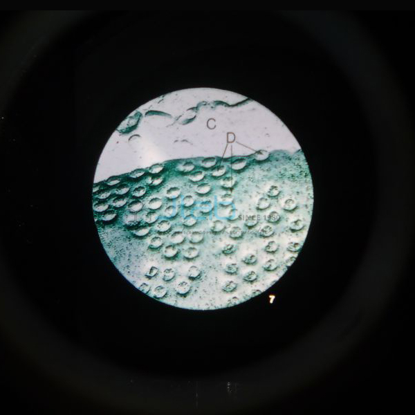
Visualizing Mitosis Poster (code EL-CE-6760) is a biology poster that lets students review and compare the steps of plant and animal mitosis. Each mitotic stage is drawn as a detailed illustration and paired with close-up photomicrographs of an onion root tip and a whitefish blastula undergoing mitosis, linking the diagram to what is seen down a microscope. JLab (Jain Laboratory Instruments Pvt Ltd, Ambala, India) supplies it to schools, colleges and dealers.
Specifications
| Parameter | Detail |
|---|---|
| Format | Poster with detailed illustrations of each mitotic stage |
| Photomicrographs | Corresponding close-ups of an onion root tip and a whitefish blastula undergoing mitosis |
| Compares | The steps of mitosis in plants and in animals |
| Quality check | Each poster is visually inspected before dispatch |
| Pack quantity | Each (sold individually) |
| Minimum order quantity | 10 units |
| Dispatch time | Usually 7–10 working days |
| Product code | EL-CE-6760 |
| Supplied by | Jain Laboratory Instruments Pvt Ltd (JLab), Ambala, India |
How Does It Compare with JLab Mitosis Charts?
| Item | Coverage | Size and finish, as listed |
|---|---|---|
| This poster (EL-CE-6760) | Plant and animal mitosis together, with photomicrographs | Not stated |
| Plant Cell Mitosis Chart | Mitosis in plant cells | 58 x 90 cm; laminated on both sides, plastic rollers |
| Animal Cell Mitosis Chart | Mitosis in animal cells | 58 x 90 cm; laminated on both sides, plastic rollers |
Choose the poster when plant and animal mitosis should be compared on one display with real photomicrographs; the two charts treat each kind of cell separately.
What Is the Mitosis Poster Used For?
The onion root tip and the whitefish blastula are the classic specimens for mitosis: the root tip grows quickly from a zone of dividing cells, and the blastula is an early fish embryo whose cells divide again and again. Pairing each illustrated stage with a photomicrograph shows students what prophase, metaphase, anaphase and telophase really look like before they search for them on a slide. The plant and animal sequences side by side highlight the differences examiners ask about — animal cells have centrioles and asters and pinch in two with a furrow, while plant cells have no centrioles and build a cell plate between the daughter nuclei. The poster fits the class 11 chapter on the cell cycle and cell division, the onion root tip practical, and first-year college biology. Hang it near the microscopes during practical sessions.
Frequently Asked Questions
Which specimens appear in the photomicrographs?
An onion root tip, for plant mitosis, and a whitefish blastula, for animal mitosis, each shown undergoing mitosis next to the matching illustrated stage.
What size is the Visualizing Mitosis Poster?
The listing does not state the size or finish; ask JLab to confirm them in your quotation.
Is there a matching meiosis poster?
Yes, the Visualising Meiosis Poster compares plant and animal meiosis in the same way.
Who supplies this mitosis poster?
Jain Laboratory Instruments Pvt Ltd (JLab), Ambala, Haryana, India, which quotes for it directly.
Supplier
JLab is the trading name of Jain Laboratory Instruments Pvt Ltd, incorporated in 2017 and based on Hargolal Road, Ambala, Haryana. The company produces science lab equipment and resells selected posters and teaching aids from other publishers, including this one, so that schools and dealers can buy apparatus and display material in one order; each poster is visually inspected before dispatch. It holds GSTIN 06AADCJ9848D1ZE and Importer-Exporter Code 3317504552. JLab exports worldwide and ships to buyers in every region — for example Cyprus in Southern Europe, Turkmenistan in Central Asia, Switzerland in Western Europe, Fiji in Oceania and DR Congo in Central Africa.
Related Mitosis Resources
- Visualising Meiosis Poster — the companion poster on plant and animal meiosis.
- Mitosis Prepared Slide — onion root and horse ascarid egg slides to find the stages under a microscope.
- Mitosis Model — enlarged views of five phases of mitosis.
- Mitosis Simulation Investigation — students model each stage with pop-bead chromosomes.
Browse Cell Equipments and all Biology Lab Equipment.
Request a Quote
JLab quotes directly. Use the Add To Cart enquiry button on this page, or send the number of posters (minimum 10 units), any companion posters or slides, and your delivery location through the contact page.
Last updated: 4 October 2026