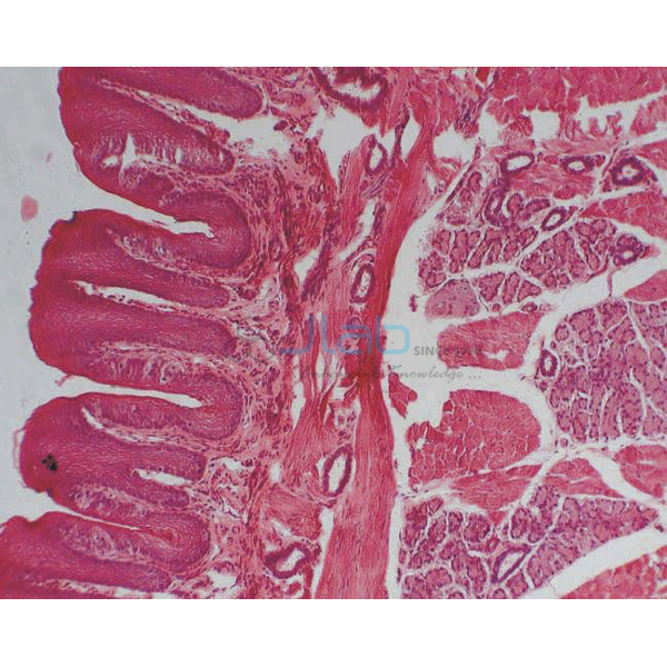

Microslide Basic Human Histology Manufacturer,Supplier and Exporter in India

Product Code : EL-CE-6733
Jlab Export is a leading Microslide Basic Human Histology Manufacturer,and suppliers in India
Microslide Basic Human Histology (code EL-CE-6733) is a Microslide set of 8 related 35 mm images, photographed through a microscope, that introduces the basic tissues of the human body: fat cells, ciliated epithelial cells, a stained bone cross-section, cartilage, stained nerve cells, striated muscle cells, and white and red blood cells. Arrows and callouts point out the key features. JLab (Jain Laboratory Instruments Pvt Ltd, Ambala, India) supplies it to schools, colleges and dealers.
Specifications
| Parameter | Detail |
|---|---|
| Brand | Microslide |
| Format | Set of 8 related 35 mm images photographed through a microscope |
| Images | Fat cells; epithelial tissue / ciliated epithelial cell; bone XS (cross-section), stained; cartilage; nerve cells, stained; striated muscle cells; white blood cells; red blood cells |
| Labelling | Arrows and callouts help students locate the important features being studied |
| Text folder | Detailed text folder designed to stimulate, inform and question students; a pocket stores the Microslides |
| Viewing | Viewed through a Microslide Viewer, available separately |
| Quality check | Each set is visually inspected before dispatch |
| Pack quantity | Each (one set) |
| Minimum order quantity | 10 units |
| Dispatch time | Usually 7–10 working days |
| Product code | EL-CE-6733 |
| Supplied by | Jain Laboratory Instruments Pvt Ltd (JLab), Ambala, India |
How Does It Compare with Other Microslide Tissue Sets?
| Set | Topic | Images listed |
|---|---|---|
| This set (EL-CE-6733) | Basic human histology | Fat cells, ciliated epithelial cells, stained bone and nerve cells, cartilage, striated muscle, white and red blood cells |
| Microslide Animal Tissue Epithelium & Cartilage | Epithelium and cartilage | Trachea at two magnifications, cartilage, oesophagus epithelium, ciliated columnar, simple cuboidal and simple squamous epithelium, urinary bladder |
| Microslide Animal Tissue Set 2 | Muscle, bone and connective tissue | Involuntary, heart and voluntary muscle cells, blood, adipose tissue, spinal cord, areolar connective tissue, bone |
This set gives one example from each main tissue group in a single folder, which suits a short introduction or revision. For a fuller unit, the two animal tissue sets divide the topic between lining tissues and muscle and connective tissues.
What Is the Basic Human Histology Microslide Used For?
The set introduces the four basic tissue types — epithelial, connective, muscle and nervous — for the tissues chapter of class 9 science, the animal tissues topic of class 11 biology, and human biology, nursing and paramedical foundation courses. Students can compare the many cell forms within connective tissue (fat cells, bone, cartilage and blood), find the cilia on the epithelial cells, trace the long processes of the nerve cells and the stripes of the striated muscle, and set white blood cells, which have nuclei and help defend the body, against mature red blood cells, which have none.
Because the images are seen through a Microslide Viewer, the lesson needs no microscope set-up, and the arrows and callouts guide students to each feature. The text folder adds reading and questions for follow-up, and its pocket keeps the Microslides together.
Frequently Asked Questions
Which images are in Microslide Basic Human Histology?
Fat cells, epithelial tissue with a ciliated epithelial cell, a stained cross-section (XS) of bone, cartilage, stained nerve cells, striated muscle cells, white blood cells and red blood cells — eight images.
What does “XS” mean on the bone image?
XS stands for cross-section: the bone was cut across its length, which shows its structure end-on, and the section was stained to make the features visible.
Do I need a microscope?
No. The Microslides are viewed through a Microslide Viewer, a separate item that needs no batteries — a white reflector lights the image in a normal classroom — and focuses with a rack and pinion.
Who supplies this Microslide set?
JLab, the trading name of Jain Laboratory Instruments Pvt Ltd in Ambala, India, supplies this Microslide-brand set and quotes for it directly.
Supplier
JLab is the trading name of Jain Laboratory Instruments Pvt Ltd, a company in Ambala, Haryana, India, incorporated in 2017 and based on Hargolal Road, that supplies laboratory and science teaching equipment. It offers Microslide sets alongside its own laboratory and biology range, so a school or dealer can place one order for viewers, Microslides and lab equipment, and every set is visually inspected before dispatch. JLab exports worldwide and ships to buyers in every region — for example Japan in East Asia, Estonia in Northern Europe, Indonesia in Southeast Asia, Guatemala in Central America and Yemen in the Middle East.
Related Microslides and Slide Sets
- Microslide Viewer — the battery-free viewer for all Microslide sets.
- Microslide Animal Tissue Epithelium & Cartilage — eight images of epithelial tissues and cartilage.
- Microslide Animal Tissue Set 2 — muscle, bone and connective tissue images.
- Human Histology Slide Set of 25 — glass microscope slides of human tissues for microscope practicals.
See all items in the Cell Equipments range or the full Biology Lab Equipment range.
Request a Quote
Quotes come direct from JLab. Use the Add To Cart enquiry button on this page, or send the number of sets (minimum 10 units), how many Microslide Viewers you need, and your delivery location through the contact page.
Last updated: 4 October 2026