
Microslide Cell Membrane Manufacturer,Supplier and Exporter in India

Product Code : EL-CE-6734
Jlab Export is a leading Microslide Cell Membrane Manufacturer,and suppliers in India
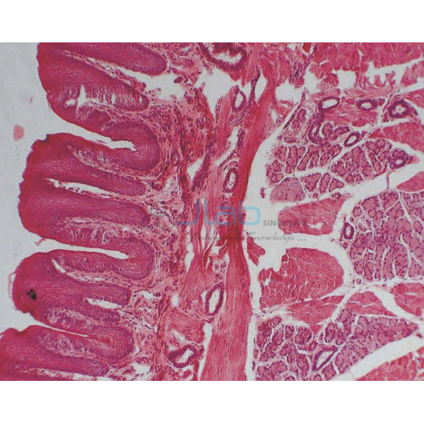
Microslide Cell Membrane (code EL-CE-6734) is a Microslide lesson set on the cell membrane: related 35 mm images, photographed through a microscope, of Paramecium cilia, blood cell structures and functions, epithelial cells, the cell membrane itself, onion skin cells and white blood cell recognition, with arrows and callouts that show students where to look. JLab (Jain Laboratory Instruments Pvt Ltd, Ambala, India) supplies it to schools, colleges and dealers.
Specifications
| Parameter | Detail |
|---|---|
| Brand | Microslide |
| Format | Microslide set (Microslides are sets of 8 related 35 mm images photographed through a microscope) |
| Images listed | Paramecium (dying) cilia; blood cells (structures); epithelial cells; cell membrane; blood cells (functions); onion skin cells; white blood cells (recognition) |
| Labelling | Arrows and callouts help students locate the important features being studied |
| Text folder | Detailed text folder designed to stimulate, inform and question students, with a pocket for the Microslides |
| Viewing | Through a Microslide Viewer, quoted as a separate item |
| Quality check | Each set is visually inspected before dispatch |
| Pack quantity | Each (one set) |
| Minimum order quantity | 10 units |
| Dispatch time | Usually 7–10 working days |
| Product code | EL-CE-6734 |
| Supplied by | Jain Laboratory Instruments Pvt Ltd (JLab), Ambala, India |
How Does It Compare with Other Microslide Cell Sets?
| Set | Topic | Images listed |
|---|---|---|
| This set (EL-CE-6734) | The cell membrane | Paramecium cilia, blood cell structures and functions, epithelial cells, cell membrane, onion skin cells, white blood cell recognition |
| Microslide the Cell Nucleus | The cell nucleus | Human blood cells, voluntary muscle cells, animal and plant cell nuclei, nuclear reproduction, endoplasmic reticulum, nuclear membrane and its surface |
| The Cell Structure Microslide | Cells across living things | Green leaf, cheek cells, cork, onion skin, blood cells, a virus, nerve cells and bacteria |
| Ultra Structure of Animal Cells Microslide | Organelles of animal cells | Golgi body and vacuole, gland cells, plasma cell, mitochondrion, centriole, lamp-brush chromosomes, nerve cell membrane, muscle cell fibrils |
Choose the cell membrane set when the lesson is about the boundary of the cell and what it does. The cell structure set is a first look at whole cells, the nucleus set focuses on the control centre, and the ultra-structure set goes into organelles, so together they cover a full cell biology unit.
What Is the Cell Membrane Microslide Used For?
The cell membrane is the thin, flexible boundary of every cell, and it is selectively permeable — it controls which substances enter and leave. This Microslide set supports that topic in class 9 science (the fundamental unit of life) and class 11 biology, and in introductory college cell biology. Onion skin cells let students tell the membrane apart from the cell wall of a plant cell; blood cells show how membrane shape suits function; the white blood cell images introduce recognition, the way immune cells identify what does not belong in the body; and the cilia of Paramecium show the membrane extended into tiny hair-like structures that the organism uses to move.
The images are viewed in a Microslide Viewer, so the class can study prepared views without microscope set-up while the arrows and callouts show what to find. The text folder supplies reading and questions, and its pocket keeps the set together.
Frequently Asked Questions
Which images does Microslide Cell Membrane include?
The listing names Paramecium (dying) cilia, blood cells (structures), epithelial cells, the cell membrane, blood cells (functions), onion skin cells and white blood cells (recognition). Microslides are sets of 8 images; ask JLab to confirm the full image list with your quotation.
What else do I need?
A Microslide Viewer to look at the images. It is a separate item, needs no batteries thanks to its white reflector, and has rack-and-pinion focusing.
Who supplies Microslide Cell Membrane?
JLab (Jain Laboratory Instruments Pvt Ltd, Ambala, India) supplies this Microslide-brand set and quotes for it directly.
Supplier
JLab is the trading name of Jain Laboratory Instruments Pvt Ltd, a laboratory and educational equipment company in India, incorporated in 2017 and based on Hargolal Road, Ambala, Haryana. Alongside its own range of science and laboratory equipment it supplies Microslide sets like this one, which lets schools, colleges and dealers source viewers, Microslides and lab apparatus together; each set is visually inspected before dispatch. JLab exports worldwide and ships to buyers in every region — for example Hong Kong in East Asia, the USA in North America, North Macedonia in Southern Europe, Belgium in Western Europe and Malawi in East Africa.
Related Microslides and Cell Models
- Microslide Viewer — battery-free viewer with rack-and-pinion focusing.
- The Cell Structure Microslide — whole cells from leaf, cork and onion skin to bacteria and a virus.
- Microslide the Cell Nucleus — the nucleus, its membrane and nuclear reproduction.
- Ultra Structure of Animal Cells Microslide — Golgi body, mitochondrion, centriole and other organelles.
- Inflatable Animal Cell Model — a 600 mm classroom model for reviewing cell parts.
See all items in the Cell Equipments range or the full Biology Lab Equipment range.
Request a Quote
Quotes come direct from JLab. Use the Add To Cart enquiry button on this page, or send the number of sets (minimum 10 units), any viewers or other Microslide sets you need, and your delivery location through the contact page.
Last updated: 4 October 2026