
Skin Microslide Manufacturer,Supplier and Exporter in India
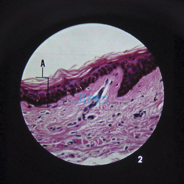
Product Code : EL-HBE-6973
Jlab Export is a leading Skin Microslide Manufacturer,and suppliers in India
Skin Microslide (code EL-HBE-6973) is a Microslide set of eight related 35 mm images, photographed through a microscope, on the structure of skin — its blood supply, sweat glands, hair and fingernail, thick and thin skin from the sole of the foot and the chest, and the mucus and poison glands of frog skin for comparison. A detailed text folder accompanies the images. JLab (Jain Laboratory Instruments Pvt Ltd, Ambala, India) supplies it to schools, colleges and dealers.
Specifications
| Parameter | Detail |
|---|---|
| Brand | Microslide |
| Format | Set of eight related 35 mm images photographed through a microscope |
| Images (as listed) | Blood supply; sweat glands; sole of the foot; fingernail; skin of the chest; hair; frog mucus; frog poison glands |
| Labelling | Arrows and callouts that help students locate the features being studied |
| Text folder | Detailed text designed to stimulate, inform and question students about the topic, with a pocket for storing the Microslide |
| Viewer | Made to be viewed through a Microslide Viewer, listed separately |
| Quality check | Each set is visually inspected before dispatch |
| Pack quantity | Each (one set) |
| Minimum order quantity | 10 units |
| Dispatch time | Usually 7–10 working days |
| Product code | EL-HBE-6973 |
| Supplied by | Jain Laboratory Instruments Pvt Ltd (JLab), Ambala, India |
What Is the Skin Microslide Used For?
The eight views take a class through what skin is made of and what it does. Sweat glands and the blood supply are the two structures the body relies on to control its temperature: sweat cools the surface as it evaporates, and blood vessels near the surface widen to lose heat or narrow to keep it. The sole of the foot and the skin of the chest show how the thickness of the outer layer changes from one part of the body to another, while the hair and fingernail images show structures that grow from the skin and are made largely of keratin. The two frog images finish the set with a contrast: amphibian skin is kept moist by mucus glands, which matters because frogs exchange gases through their skin, and poison glands help protect them from predators.
These topics appear in class 11 biology, where the frog’s moist skin and the part sweat glands play in excretion are studied, and in introductory histology for college zoology, physiology and nursing students. In a typical lesson students look at each image in a Microslide Viewer, use the arrows and callouts to find the feature named, and then answer the questions in the text folder in class or as homework. Between lessons the Microslide goes back into the folder pocket.
What to Specify When Ordering
- Number of sets — the minimum order is 10 units, for example one set per bench or group.
- Microslide Viewers — the images are made to be seen through one, so add viewers if the lab has none.
- Three-dimensional follow-up — a skin section model or block model can be added to the same quotation.
Frequently Asked Questions
What does the Skin Microslide show?
Eight images: blood supply, sweat glands, the sole of the foot, a fingernail, skin of the chest, hair, frog mucus and frog poison glands, each photographed through a microscope.
Why does a skin set include frog skin?
For comparison. Frog skin stays moist with mucus because frogs exchange gases through it, and its poison glands are a defence, whereas human skin has a dry, keratinised surface and uses sweat glands and blood flow to control body temperature.
Do I need a Microslide Viewer?
Yes. The set is made to be viewed through a Microslide Viewer, which JLab lists as a separate item. Say in your enquiry whether you need viewers as well.
What is in the text folder?
Detailed text written to stimulate, inform and question students about the skin, plus a pocket in which the Microslide is stored.
Who supplies the Skin Microslide?
JLab — Jain Laboratory Instruments Pvt Ltd of Ambala, Haryana, India. It is a Microslide product that JLab resells and quotes for directly, alongside its own human biology range.
Supplier
JLab is the trading name of Jain Laboratory Instruments Pvt Ltd, a company incorporated in 2017 and based on Hargolal Road, Ambala, Haryana, that equips school and college science laboratories. Microslide sets are among the third-party teaching resources it resells next to its own biology models and charts, so a school can buy skin models, wall charts and photomicrograph sets from one source; every set is visually inspected before dispatch. JLab exports worldwide, shipping to buyers in every region — Iceland in Northern Europe, Eswatini in Southern Africa, Uzbekistan in Central Asia, Nigeria in West Africa and Qatar in the Middle East, for example.
Related Skin Biology Resources
- Microslide Viewer — designed for viewing Microslides; a white reflector replaces batteries.
- Skin Section Model — shows the layers of the skin with hair follicles, sweat and sebaceous glands, receptors, nerves and vessels.
- Human Skin Model — a labelled block model on a board showing the skin layers and the structures within them.
- Human Skin Chart — a wall chart of the skin’s layers, glands, hair follicles and sense receptors.
- The Reflex Arc Microslide — a Microslide set that begins with the Pacinian corpuscle, a pressure receptor in the skin.
Browse the Human Biology Equipments category or all Biology Lab Equipment.
Request a Quote
Quotes come direct from JLab. Use the Add To Cart enquiry button on this page, or send the number of Skin Microslide sets (minimum 10 units), any viewers or skin models to include, and the delivery location through the contact page.
Last updated: 5 October 2026