
Micro Anatomy of Eye Manufacturer,Supplier and Exporter in India

Product Code : EL-AM-6047
Jlab Export is a leading Micro Anatomy of Eye Manufacturer,and suppliers in India
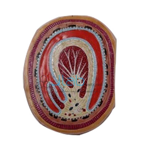
Micro Anatomy of Eye (code EL-AM-6047) is a histology teaching model of the microscopic structure of the retina, shown together with the choroid and the sclera that lie outside it. JLab (Jain Laboratory Instruments Pvt Ltd, Ambala, India) supplies it to schools, colleges and dealers.
Specifications
| Parameter | Detail |
|---|---|
| Model type | Micro anatomy (histology) model of the eye |
| Shows | The microscopic structure of the retina with the choroid and sclera |
| Left side | Block-like and layered: the complete structure of the retina, including the supplying vascular layer and parts of the sclera, as seen under a light microscope |
| Right side | Sectional enlargement showing the microscopic structure of the photoreceptor and of the cells of the pigmented layer |
| Quality check | Each model is visually inspected before dispatch |
| Pack quantity | Each (sold individually) |
| Minimum order quantity | 10 units |
| Dispatch time | Usually 7–10 working days |
| Product code | EL-AM-6047 |
| Supplied by | Jain Laboratory Instruments Pvt Ltd (JLab), Ambala, India |
What Is the Micro Anatomy of Eye Model Used For?
It teaches the layered structure of the retina and how the retina lies against the vascular choroid and the tough sclera. The photoreceptors and the pigmented layer, shown enlarged, form the outer part of the retina, where light is actually detected.
Frequently Asked Questions
What does the pigmented layer of the retina do?
This single layer of cells absorbs stray light, recycles the visual pigment used by the photoreceptors and supports and nourishes them, as they lie directly against it. The choroid behind it carries the blood supply for the outer retina.
How does it differ from the Giant Eye Model?
The Giant Eye Model is a model of the whole eyeball whose parts detach to show the three coats of the eye wall, the lens and the vitreous body. This model shows the retina, with the choroid and sclera, at microscopic level.
What details does an export quotation need?
The number of models, the destination country and port or city, the delivery terms you would like (for example FOB or CIF), any packing or labelling instructions, and a tender reference where one applies.
Who supplies the Micro Anatomy of Eye model?
JLab, the trading name of Jain Laboratory Instruments Pvt Ltd in Ambala, Haryana, India, supplies it and quotes for it directly.
Supplier
Jain Laboratory Instruments Pvt Ltd, trading as JLab, is a laboratory and educational equipment company incorporated in 2017 and located on Hargolal Road, Ambala, Haryana. Schools, colleges, dealers and importers can order this eye histology model from JLab together with its own anatomy models; every piece is looked over before dispatch and repeat orders are handled directly. JLab exports worldwide and ships to buyers in every region — for example Japan in East Asia, the UK in Western Europe, Algeria in North Africa, Iraq in the Middle East and Guatemala in Central America and the Caribbean. For tenders, see the JLab lab tenders page.
Related Eye Models
- Giant Eye Model — the whole eyeball with detachable parts.
- Human Eye 5 Times — an enlarged eye that opens in layers, the upper half of the sclera coming off to expose the choroid.
- Functional Eye Model — a working eye with a flexible lens that focuses an image on the retina.
- L.S. Insect Eye — an enlarged model of the histological microstructure of an insect eye, mounted on a stand.
- Micro Anatomy, Muscle Fiber — a skeletal muscle fiber and its neuromuscular end plate, greatly magnified.
Find more in the Anatomical Models category and the wider anatomical models range.
Request a Quote
For a quotation, press Add To Cart to send an enquiry or use the contact page, and tell us the quantity and the delivery city and country.
Last updated: 22 October 2026