
Animal Cell Organelles Chart Manufacturer,Supplier and Exporter in India
Product Code : EL-ZC-9310
Jlab Export is a leading Animal Cell Organelles Chart Manufacturer,and suppliers in India
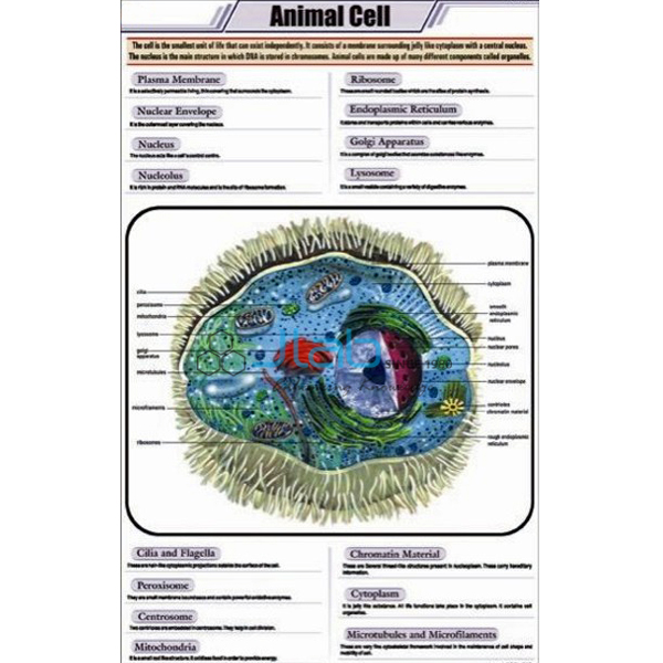
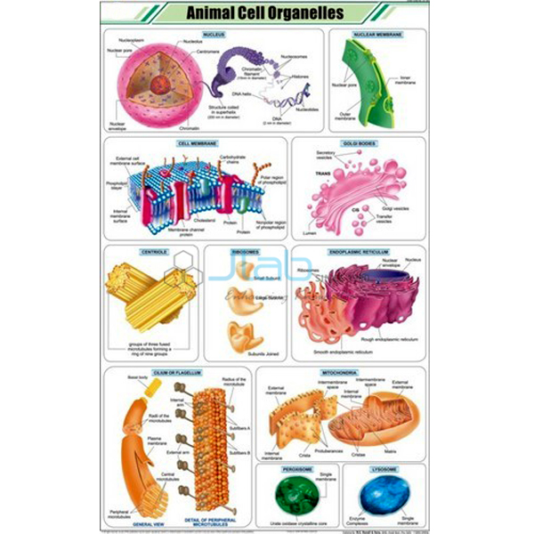
Animal Cell Organelles Chart (code EL-ZC-9310) is a 58 x 90 cm biology wall chart on the organelles of the animal cell, used to teach their structure and functions in higher-secondary and college cell biology. It is printed in full colour on art paper, laminated on both sides with thick plastic film and fitted with plastic rollers, with English text only. JLab (Jain Laboratory Instruments Pvt Ltd, Ambala, India) supplies it to schools, colleges and dealers.
Specifications
| Parameter | Detail |
|---|---|
| Chart topic | Organelles of the animal cell |
| Size | 58 x 90 cm (about 22.8 x 35.4 in) |
| Printing | Full colour on art paper |
| Lamination | Both sides, thick plastic film |
| Fittings | Plastic rollers |
| Language | English only |
| Quality check | Each chart is visually inspected before dispatch |
| Pack quantity | Each (sold individually) |
| Minimum order quantity | 10 units |
| Dispatch time | Usually 7–10 working days |
| Product code | EL-ZC-9310 |
| Supplied by | Jain Laboratory Instruments Pvt Ltd (JLab), Ambala, India |
What Is the Animal Cell Organelles Chart Used For?
Once students know what an animal cell looks like, the next question is what each part does. The class 11 chapter on the cell as the unit of life works through the organelles one by one: the nucleus, holding the cell's DNA inside a double envelope with pores; mitochondria, where aerobic respiration releases energy as ATP; ribosomes, which build proteins, some of them attached to the rough endoplasmic reticulum, while the smooth endoplasmic reticulum makes lipids; the Golgi apparatus, which modifies and packages materials for secretion; lysosomes, the membrane sacs of digestive enzymes; and the centrosome, whose centrioles organise the spindle in cell division and form the basal bodies of cilia and flagella. The cytoskeleton of microtubules and microfilaments gives the cell its shape and lets it move.
Most of these structures are visible only with an electron microscope, so an organelle chart is how a class sees them in detail. Teachers use it for the structure-and-function tables that appear in tests, and it pairs well with a general animal cell chart that shows the cell as a whole. Wipe the laminated face with a damp cloth and roll the chart on its rollers for storage. The listing does not describe the artwork, so ask JLab for a larger image if you need to check which organelles are drawn.
What to Specify When Ordering
- How many charts — the minimum is 10 units, each one supplied individually.
- Whether you also need the general animal cell chart or the plant cell organelles chart for comparison lessons.
- Any cell models to quote with the charts.
- Where the order is going — city and country, and for export the port and the delivery terms (for example FOB or CIF).
Frequently Asked Questions
What is this chart used to teach?
It is used to teach the structure and function of the organelles inside an animal cell. The listing does not describe the artwork; the product photograph shows the chart, and JLab can provide a larger image before you order.
What are the chart's size and finish?
It is 58 x 90 cm (about 22.8 x 35.4 inches), printed in full colour on art paper and laminated on both sides with thick plastic film, with plastic rollers for hanging.
Which organelle releases energy for the cell?
The mitochondrion. Its folded inner membrane carries out the final stages of aerobic respiration, which release energy from food and store it as ATP, the molecule cells use to power their work.
Can these organelles be seen with a school microscope?
Only some of them. A light microscope shows the nucleus clearly, but most other organelles are too small and were described using the electron microscope, which is why diagrams and charts are used to teach them.
Who supplies this organelles chart?
Jain Laboratory Instruments Pvt Ltd, trading as JLab, from Ambala, Haryana, India, supplies the chart and quotes for it directly.
Supplier
JLab is the trading name of Jain Laboratory Instruments Pvt Ltd, incorporated in 2017 and based on Hargolal Road, Ambala, Haryana, a supplier of laboratory and teaching equipment to education buyers. In addition to its own equipment range it resells teaching charts such as this one to schools, colleges, dealers and importers; every chart is visually checked before packing, and repeat orders are dealt with by JLab itself. JLab exports worldwide and supplies buyers in every region, for example Norway in Northern Europe, Namibia in Southern Africa, Madagascar in East Africa, Guatemala in Central America and the Caribbean and Hungary in Eastern Europe. For purchases through tenders, see the JLab lab tenders page.
Related Cell Biology Charts and Models
- Animal Cell Chart — the companion chart in this series showing the whole animal cell.
- Plant Cell Organelles Chart — the plant-cell counterpart from the botany chart series.
- Animal Cell — an enlarged model showing organelles such as the nucleus, endoplasmic reticulum, mitochondria and ribosomes.
- Animal Cell Model — a model that also shows centrioles, lysosomes and fat vacuoles.
- Animal Cell Mitosis Chart — for the lesson on how the cell and its nucleus divide.
See all titles in the Zoology Chart range or the full Charts range.
Request a Quote
For a quotation from JLab, use the Add To Cart enquiry button on this page or send the quantity (minimum 10 units), any companion charts or models and your delivery location through the contact page.
Last updated: 3 October 2026