
You have searched for "Human"

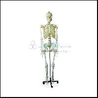
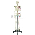
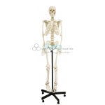
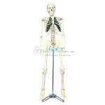
Human Skeleton Tall 180 cm
Product Code : JL-HM-133
Basics of Human Anatomy. Offered skeleton life is fabricated using supreme class raw PVC plastic that is sourced from the authentic vendor of the market, in compliance with international quality standards. View Details
Basics of Human Anatomy. Offered skeleton life is fabricated using supreme class raw PVC plastic that is sourced from the authentic vendor of the market, in compliance with international quality standards. View Details
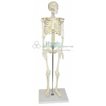
Human Skeleton Medium Tall 85cm
Product Code : JL-HM-134
Skeleton with full natural movement and a user-friendly personality that will encourage children to learn the names of the bones. View Details
Skeleton with full natural movement and a user-friendly personality that will encourage children to learn the names of the bones. View Details
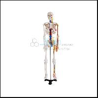
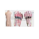
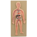
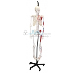
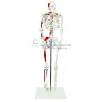
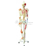
Human Skeleton with Nerves and Blood Vessels Tall 85cm
Product Code : JL-HM-135
This model depicts the position, course and distribution of main arteries and peripheral nerves of the human body. It may be employed as a visual aid in the instruction of anatomy to students of medicine View Details
This model depicts the position, course and distribution of main arteries and peripheral nerves of the human body. It may be employed as a visual aid in the instruction of anatomy to students of medicine View Details

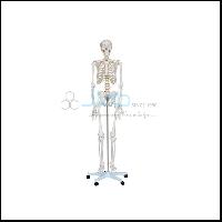
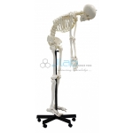
Human Skeleton Life - Size Tall 170cm
Product Code : JL-HM-136
We are offering them a commendable range of Life Size Skeleton 170 CM Tall Models. It is sourced by our diligent professionals so as to ensure its quality and prolonged service life. View Details
We are offering them a commendable range of Life Size Skeleton 170 CM Tall Models. It is sourced by our diligent professionals so as to ensure its quality and prolonged service life. View Details
Mini Human Skeleton 42cm
Product Code : JL-HM-137
Our clients can avail from us superior quality range of Mini Human Skeleton that is available at the most reasonable prices. View Details
Our clients can avail from us superior quality range of Mini Human Skeleton that is available at the most reasonable prices. View Details
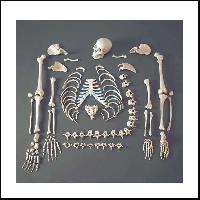
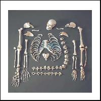
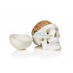
Disarticulated Human Skeleton 200 Bones
Product Code : JL-HM-138
This human skeleton is designed in compliance with international quality standards utilizing high grade material of standard quality and cutting-edge technology. View Details
This human skeleton is designed in compliance with international quality standards utilizing high grade material of standard quality and cutting-edge technology. View Details
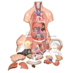
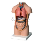
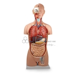
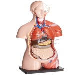
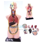
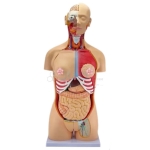
Human Torso 15 Parts Tall 26cm
Product Code : JL-AM-163
This most popular educational torso features: Torso, brain (2 parts), cut calvarium, heart, lung (4 parts), stomach, diaphragm, liver, heart, trachea & esophagus & aorts, pancreas and spleen, intestine etc. View Details
This most popular educational torso features: Torso, brain (2 parts), cut calvarium, heart, lung (4 parts), stomach, diaphragm, liver, heart, trachea & esophagus & aorts, pancreas and spleen, intestine etc. View Details
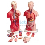
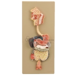
Unisex Torso and Human Torso
Product Code : JL-AM-164
This outstanding torso features an exposed spine with removable vertebrae and spinal cord segments, a female breast plate and interchangeable male and female genitalia. View Details
This outstanding torso features an exposed spine with removable vertebrae and spinal cord segments, a female breast plate and interchangeable male and female genitalia. View Details
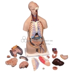
Human Torso 90cms
Product Code : JL-AM-165
This full size high quality muscular figure represents a wide variety of human anatomical structures with accurate details. View Details
This full size high quality muscular figure represents a wide variety of human anatomical structures with accurate details. View Details
Human Torso Vertical Shears
Product Code : JL-AM-167
Human Torso 90cms Vertical Shears 10 pcs View Details
Human Torso 90cms Vertical Shears 10 pcs View Details
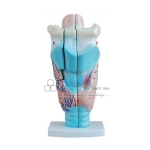
Magnified Human Larynx Model
Product Code : JL-AM-179
A functional model that demonstrates movements of the epiglottis and cartilages in the voice box. View Details
A functional model that demonstrates movements of the epiglottis and cartilages in the voice box. View Details
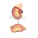
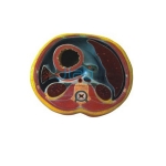
Human Kidney with Adrenal Gland
Product Code : JL-AM-181
The detailed life-size model features the kidney, adrenal gland, renal and adrenal vessels and upper portion of the urethra. View Details
The detailed life-size model features the kidney, adrenal gland, renal and adrenal vessels and upper portion of the urethra. View Details
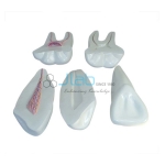
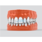
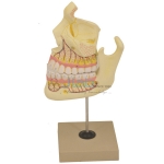
Expansion Model of Human Teeth
Product Code : JL-AM-190
Our rise to recognition in this industry has been primarily triggered by the shooting popularity of our offered range of Expansion Model of Human Teeth. View Details
Our rise to recognition in this industry has been primarily triggered by the shooting popularity of our offered range of Expansion Model of Human Teeth. View Details
Human Body Anatomy Model
Product Code : JL-AM-197
Being a customer oriented enter prise, we are engaged in providing a wide array of Human Full Body Anatomy Model. View Details
Being a customer oriented enter prise, we are engaged in providing a wide array of Human Full Body Anatomy Model. View Details
Transparent Human Skeleton Tall 85cm
Product Code : JL-AM-201
Dissectible into 2 parts, removable from base, shows internal and external anatomy including valves, cardiac chambers and pulmonary and systemic vascular structures. View Details
Dissectible into 2 parts, removable from base, shows internal and external anatomy including valves, cardiac chambers and pulmonary and systemic vascular structures. View Details
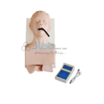
Human Trachea Intubation Model
Product Code : JL-AM-202
This model is designed as a teaching model for training the medical students. Basic clinic nurses and first aid people to demonstrate and practice trachea intubation through mouth. View Details
This model is designed as a teaching model for training the medical students. Basic clinic nurses and first aid people to demonstrate and practice trachea intubation through mouth. View Details
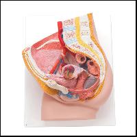
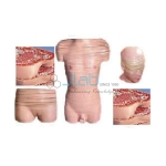
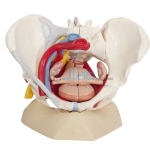
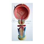
Human Female Pelvis Section
Product Code : JL-NTM-218
The model is a median section shows female genital organs with bladder and rectum. The abdominal and pelvis muscles are shown detailed. View Details
The model is a median section shows female genital organs with bladder and rectum. The abdominal and pelvis muscles are shown detailed. View Details
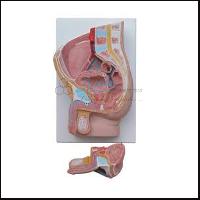
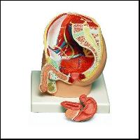
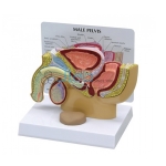
Human Male Pelvis Section
Product Code : JL-NTM-219
This median section model shows the normal position of male genital organs with bladder and rectum in the male pelvis. Made of PVC. View Details
This median section model shows the normal position of male genital organs with bladder and rectum in the male pelvis. Made of PVC. View Details
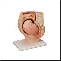
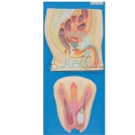
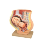
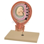
Human Female Pregnant Pelvis Section with Fetus
Product Code : JL-NTM-220
Life size representation of a meidn section through the female pelvis in the 9th month of prenancy with removable fetus. View Details
Life size representation of a meidn section through the female pelvis in the 9th month of prenancy with removable fetus. View Details
Human Male Pelvis Section 2 Part
Product Code : JL-NTM-221
This median section model shows the normal position of male genital organs with bladder and rectum in the male pelvis. Dissectible into 2 parts. Packing: 4pcs/carton, 37x55x37cm, 10kgs View Details
This median section model shows the normal position of male genital organs with bladder and rectum in the male pelvis. Dissectible into 2 parts. Packing: 4pcs/carton, 37x55x37cm, 10kgs View Details
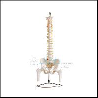
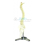
Human Vertebral Column
Product Code : JL-OM-264
This life size vertebral column of an adult human shows all the special features of vertebral, pelvis. We are the leading manufacturer of this product. This life size vertebral column of an adult human shows all the special features of vertebral, pe View Details
This life size vertebral column of an adult human shows all the special features of vertebral, pelvis. We are the leading manufacturer of this product. This life size vertebral column of an adult human shows all the special features of vertebral, pe View Details
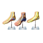
Human Foot Joint Model
Product Code : JL-OM-267
This life size foot model demonstrates whole anatomy of the foot & is useful for any one studying. The human anatomy or for doctors to explain the problems to the patients. View Details
This life size foot model demonstrates whole anatomy of the foot & is useful for any one studying. The human anatomy or for doctors to explain the problems to the patients. View Details
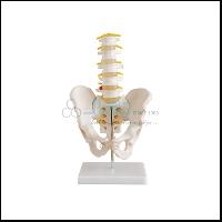
Human Male Pelvis with Lumbar Vertebrae
Product Code : JL-OM-272
Consisting of hip bone. Sacrum with coccyx and 5 lumbar vertebrae. Mounted on a base. This model shows all the features of adult human male pelvis. View Details
Consisting of hip bone. Sacrum with coccyx and 5 lumbar vertebrae. Mounted on a base. This model shows all the features of adult human male pelvis. View Details
Human Mini Spine Model
Product Code : JL-OM-273
MMini size spine model shows all 24 vertebrae with pelvis. Sacrum. View Details
MMini size spine model shows all 24 vertebrae with pelvis. Sacrum. View Details

Human Male Pelvis Model
Product Code : JL-OM-275
Human male pelvis model shows the shape. Size & other special features of adult male which make it different from female pelvis. View Details
Human male pelvis model shows the shape. Size & other special features of adult male which make it different from female pelvis. View Details
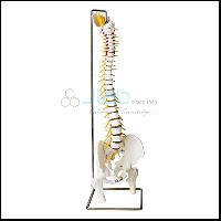
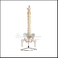
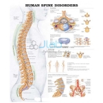
Human Spinal Column Model
Product Code : JL-OM-280
This life-size spinal column model with pelvis presents the anatomical structure of the spine with the additional pathological details of a herniated disc. View Details
This life-size spinal column model with pelvis presents the anatomical structure of the spine with the additional pathological details of a herniated disc. View Details
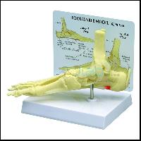
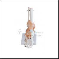
Human Foot Model
Product Code : JL-OM-281
Full size model features plantar Calcagno acicular ligament with plantar fasciitis. Foot/ankle anatomy also includes: tibia, fibula, calcaneus, calcaneal tendon, deltoid ligament, lateral ligament, plantar. View Details
Full size model features plantar Calcagno acicular ligament with plantar fasciitis. Foot/ankle anatomy also includes: tibia, fibula, calcaneus, calcaneal tendon, deltoid ligament, lateral ligament, plantar. View Details
Deluxe Human Disarticulated Skeleton
Product Code : JL-SFMS-321
Human Skeleton bones near to the original preferred by medical students and medical institutes approved by MCI. For study based all Skeleton Parts are Near to Original Skull with Mandible 3parts. View Details
Human Skeleton bones near to the original preferred by medical students and medical institutes approved by MCI. For study based all Skeleton Parts are Near to Original Skull with Mandible 3parts. View Details
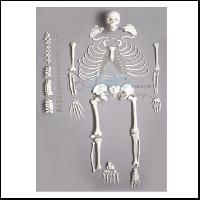
Human Skeleton Dis-articulated
Product Code : JL-SFMS-322
The identification of individual bones is easy enough for students using a fully constructed skeleton, but can your students recognize a femur on its own. View Details
The identification of individual bones is easy enough for students using a fully constructed skeleton, but can your students recognize a femur on its own. View Details
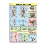
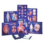
Human Skeleton Chart
Product Code : JL-HPC-2982
Printed on Synthetic in original colours Size 70 x 100 cm View Details
Printed on Synthetic in original colours Size 70 x 100 cm View Details
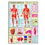
Human Muscles Chart
Product Code : JL-HPC-2983
Printed on Synthetic in original colours. Size 70 x 100 cm Available in English langauge only View Details
Printed on Synthetic in original colours. Size 70 x 100 cm Available in English langauge only View Details
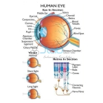
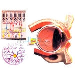
Human Eye Chart
Product Code : JL-HPC-2987
Printed on Synthetic in original colours. Size:- 70 x 100 cm View Details
Printed on Synthetic in original colours. Size:- 70 x 100 cm View Details
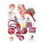
Human Ear Chart
Product Code : JL-HPC-2988
Available in English langauge only Printed on Synthetic in original colours View Details
Available in English langauge only Printed on Synthetic in original colours View Details
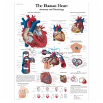
Human Heart Chart
Product Code : JL-HPC-2991
Printed on Synthetic in original colours Size:- 70 x 100 cm View Details
Printed on Synthetic in original colours Size:- 70 x 100 cm View Details
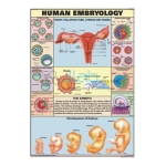
Human Embryology Chart
Product Code : JL-HPC-2994
Printed on Synthetic in original colours. Size:- 70 x 100 cm View Details
Printed on Synthetic in original colours. Size:- 70 x 100 cm View Details
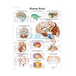
Human Brain Chart
Product Code : JL-HPC-2996
Printed on Syhnthetic paper in original colours. Chart size 70 x 100 cms View Details
Printed on Syhnthetic paper in original colours. Chart size 70 x 100 cms View Details
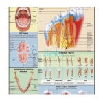
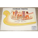
Human Teeth Chart
Product Code : JL-HPC-2997
Printed on Syhnthetic paper in original colours. Chart size 70 x 100 cms. View Details
Printed on Syhnthetic paper in original colours. Chart size 70 x 100 cms. View Details
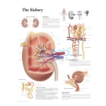
Human Kidney Chart
Product Code : JL-HPC-2999
Printed on Syhnthetic paper in original colours. Chart size:- 70 x 100 cms View Details
Printed on Syhnthetic paper in original colours. Chart size:- 70 x 100 cms View Details
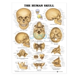
Human Skull Chart
Product Code : JL-HPC-3002
Shows anterior and lateral aspects of the skull. Illustrates base of skull (including inner surface). Sagittal section through skull, horizontal section through maxilla, mandible, coronal section through anterior skull, ethmoid bone. View Details
Shows anterior and lateral aspects of the skull. Illustrates base of skull (including inner surface). Sagittal section through skull, horizontal section through maxilla, mandible, coronal section through anterior skull, ethmoid bone. View Details
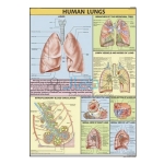
Human Lungs Chart
Product Code : JL-HPC-3005
Printed on Syhnthetic paper in original colours. Chart size 70 x 100 cms View Details
Printed on Syhnthetic paper in original colours. Chart size 70 x 100 cms View Details
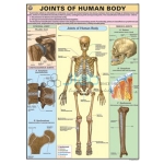
Joints of Human Body Chart
Product Code : JL-HPC-3006
Printed on Syhnthetic paper in original colours. Chart size 70 x 100 cms View Details
Printed on Syhnthetic paper in original colours. Chart size 70 x 100 cms View Details
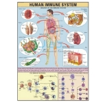
Human Immune System Chart
Product Code : JL-HPC-3007
Printed on Syhnthetic paper in original colours. Chart size 70 x 100 cms. View Details
Printed on Syhnthetic paper in original colours. Chart size 70 x 100 cms. View Details
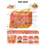
Human Skin Chart
Product Code : JL-HPC-3008
Printed on Syhnthetic paper in original colours. Chart size 70 x 100 cms. View Details
Printed on Syhnthetic paper in original colours. Chart size 70 x 100 cms. View Details
Human Spinal Cord Chart
Product Code : JL-HPC-3011
Printed on Syhnthetic paper in original colours. Chart size 70 x 100 cms. View Details
Printed on Syhnthetic paper in original colours. Chart size 70 x 100 cms. View Details

Impact of Environment Degradation on Humans Chart
Product Code : JL-MAE-3157
Explains how Ozone layer protects us, Green House effects of CO2. Available in English, Hindi & Marathi separately. View Details
Explains how Ozone layer protects us, Green House effects of CO2. Available in English, Hindi & Marathi separately. View Details
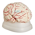
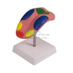
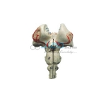
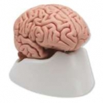
Human Brain Model
Product Code : JL-SL-4580
This Human brain model shows parts of the brain. View Details
This Human brain model shows parts of the brain. View Details
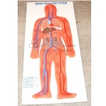
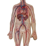
Human Circulatory System Model
Product Code : JL-SL-4581
This model shows the circulatory system of a human body. It explains which organs and veins are involved in the circulation of blood. View Details
This model shows the circulatory system of a human body. It explains which organs and veins are involved in the circulation of blood. View Details
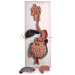
Human Digestive System Model
Product Code : JL-SL-4582
This model displays complete digestive system including nose, mouth cavity and pharynges, oesophagus, liver, pancreas, duodenum, caecum and rectum. View Details
This model displays complete digestive system including nose, mouth cavity and pharynges, oesophagus, liver, pancreas, duodenum, caecum and rectum. View Details
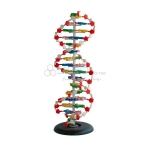
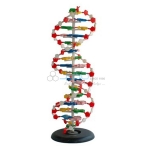
Human DNA Structure Model
Product Code : JL-SL-4583
It shows structure of DNA Molecules and their replication. View Details
It shows structure of DNA Molecules and their replication. View Details
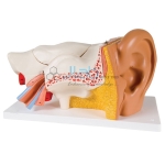
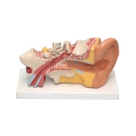
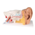
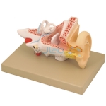
Human Ear Model
Product Code : JL-SL-4584
The model ear shows the internal structure of a human ear. The size available is standard and is well labelled. View Details
The model ear shows the internal structure of a human ear. The size available is standard and is well labelled. View Details
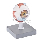
Human Eye Model
Product Code : JL-SL-4585
This model represents parts of an eye in detail with both halves of sclera with cornea, eye muscle View Details
This model represents parts of an eye in detail with both halves of sclera with cornea, eye muscle View Details
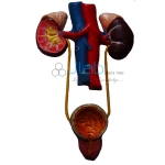
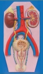
Human Excretory System Model
Product Code : JL-SL-4586
Urinary bladder, kidneys with ureter, adrenal glands, and bladder with prostate and major blood vessels are View Details
Urinary bladder, kidneys with ureter, adrenal glands, and bladder with prostate and major blood vessels are View Details

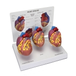
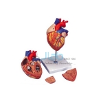
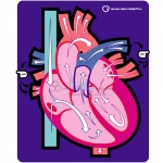
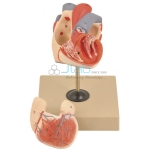
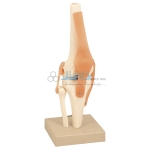
Human Heart Model
Product Code : JL-SL-4587
This heart model is designed to demonstrate the internal and external anatomy of heart showcasing valves, cardiac chamber, pulmonary and systemic vascular structures. View Details
This heart model is designed to demonstrate the internal and external anatomy of heart showcasing valves, cardiac chamber, pulmonary and systemic vascular structures. View Details
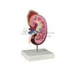
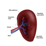
Human Kidney Model
Product Code : JL-SL-4588
This model shows the internal and external structures of a Human Kidney and Adrenal Gland along with renal View Details
This model shows the internal and external structures of a Human Kidney and Adrenal Gland along with renal View Details
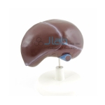
Human Liver Model
Product Code : JL-SL-4589
Well labelled model of human liver is available in standard size for educational purposes. View Details
Well labelled model of human liver is available in standard size for educational purposes. View Details
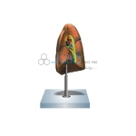
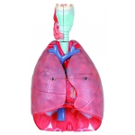
Human Lungs Model
Product Code : JL-SL-4590
Well labelled model of human lungs is available in standard size for educational purposes. View Details
Well labelled model of human lungs is available in standard size for educational purposes. View Details
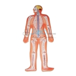
Human Nervous System Model
Product Code : JL-SL-4591
Central and peripheral nervous system are displayed in this well labelled model of human nervous system. View Details
Central and peripheral nervous system are displayed in this well labelled model of human nervous system. View Details
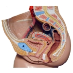
Human Pelvis Male Model
Product Code : EL-SL-4592
The male pelvis anatomy model is shown in median section One half of male genital organs with bladder is shown at the normal position in the male pelvis View Details
The male pelvis anatomy model is shown in median section One half of male genital organs with bladder is shown at the normal position in the male pelvis View Details
Human Pelvis Female Model
Product Code : JL-SL-4593
The female pelvis anatomy model is shown in median section View Details
The female pelvis anatomy model is shown in median section View Details
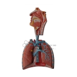
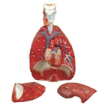
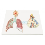
Human Respiratory System Model
Product Code : JL-SL-4594
Parts of brain in connection with respiratory system, nasal cavity, lungs, bronchial tree, etc. are represented on a cardboard base. View Details
Parts of brain in connection with respiratory system, nasal cavity, lungs, bronchial tree, etc. are represented on a cardboard base. View Details
Human Reproductive System Male Model
Product Code : JL-SL-4595
Standard sized male pelvis showing internal and external reproductive organs and other vital organs of reproductive system. View Details
Standard sized male pelvis showing internal and external reproductive organs and other vital organs of reproductive system. View Details
Human Reproductive System Female Model
Product Code : JL-SL-4596
Standard sized female pelvis showing internal and external reproductive organs and other vital organs of reproductive system. View Details
Standard sized female pelvis showing internal and external reproductive organs and other vital organs of reproductive system. View Details
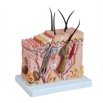
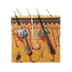
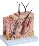
Human Skin Model
Product Code : JL-SL-4597
The model showcases the layers of skin, hair follicles, sebaceous glands, sweat glands, receptors, nerves and vessels. View Details
The model showcases the layers of skin, hair follicles, sebaceous glands, sweat glands, receptors, nerves and vessels. View Details
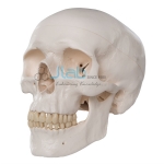
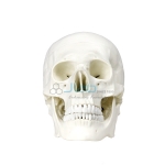
Human Skull Model
Product Code : JL-SL-4598
The model showcases the skull with teeth, upper and lower jaw made from fibre glass and clay. View Details
The model showcases the skull with teeth, upper and lower jaw made from fibre glass and clay. View Details
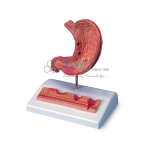
Human Stomach Model
Product Code : JL-SL-4599
This stomach pathology model demonstrates various stages of gastritis from a light gastric ulcer to a perforation. View Details
This stomach pathology model demonstrates various stages of gastritis from a light gastric ulcer to a perforation. View Details

Human Teeth Model
Product Code : JL-SL-4600
Position of different types of teeth is represented in the human teeth model. View Details
Position of different types of teeth is represented in the human teeth model. View Details
Human Tongue Model
Product Code : JL-SL-4601
The root of the tongue, epiglottis and other vital parts of the tongue are indicated in this human tongue model. View Details
The root of the tongue, epiglottis and other vital parts of the tongue are indicated in this human tongue model. View Details
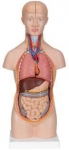
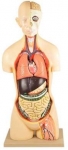
Human Torso Model (Life Size)
Product Code : JL-SL-4602
Placement of different organs like lungs, stomach, pancreas etc. are shown in this small sized human torso model. View Details
Placement of different organs like lungs, stomach, pancreas etc. are shown in this small sized human torso model. View Details

Human Torso Model (Full Size)
Product Code : JL-SL-4603
Placement of different organs like lungs, stomach, pancreas etc. are shown in this small sized human torso model. View Details
Placement of different organs like lungs, stomach, pancreas etc. are shown in this small sized human torso model. View Details
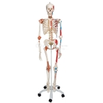
Human Skeleton Model
Product Code : JL-SL-4604
The full size model of skeleton shows articulated placement of bone and how a human body is structured. View Details
The full size model of skeleton shows articulated placement of bone and how a human body is structured. View Details
Human Artery and Vein Model
Product Code : JL-MI-4853
It is ideal for understanding the structural difference between a human artery and a vein. View Details
It is ideal for understanding the structural difference between a human artery and a vein. View Details

Human Antagonistic Muscle Model
Product Code : JL-MI-4854
All models are made from superior quality materials and are finished well complete with labels. View Details
All models are made from superior quality materials and are finished well complete with labels. View Details
Human DNA Model
Product Code : JL-MI-4856
The structure is magnified to unveil the detailed structure of the molecule. View Details
The structure is magnified to unveil the detailed structure of the molecule. View Details
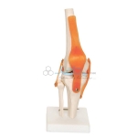
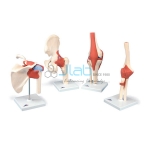
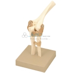
Human Knee Joint Model
Product Code : JL-MI-4867
Human knee joint demonstrates the structure of knee. View Details
Human knee joint demonstrates the structure of knee. View Details
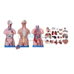
Human Torso (Unisex)
Product Code : JL-AM-5906
Outstanding torso features an exposed spine with removable vertebrae and spinal cord segments, a female breast plate and interchangeable male and female genitalia. View Details
Outstanding torso features an exposed spine with removable vertebrae and spinal cord segments, a female breast plate and interchangeable male and female genitalia. View Details
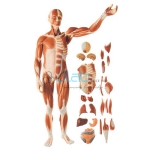
Full Size Human Body
Product Code : JL-AM-5907
Full Size Human Body showing Muscles & Organs This model consists of 27 parts, such as muscles of chest wall and abdomen, muscles of upper and lower limbs, skull, brain and viscus. View Details
Full Size Human Body showing Muscles & Organs This model consists of 27 parts, such as muscles of chest wall and abdomen, muscles of upper and lower limbs, skull, brain and viscus. View Details
Human Torso (Sexless)
Product Code : JL-AM-5908
Hand Painted in Vivid & Natural Colours 11 Dissectible Parts View Details
Hand Painted in Vivid & Natural Colours 11 Dissectible Parts View Details
Human Body Joints
Product Code : JL-AM-5914
An instructional model to illustrate abduction, adduction, anteversion, retroversion and internal / external rotation. View Details
An instructional model to illustrate abduction, adduction, anteversion, retroversion and internal / external rotation. View Details
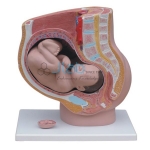
Human Female Pelvis Section
Product Code : JL-AM-5934
Shows medium section of the right half of female pelvis. A9 month fetus is removable. View Details
Shows medium section of the right half of female pelvis. A9 month fetus is removable. View Details
Human Male Pelvis Section
Product Code : JL-AM-5935
This median section model shows male genital organs with bladder and rectum in male pelvis. Made of PVC plastic. View Details
This median section model shows male genital organs with bladder and rectum in male pelvis. Made of PVC plastic. View Details
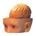
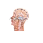
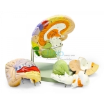
Human Head Brain
Product Code : JL-AM-5981
Science Labs shows the human brain dissectable into 2 parts, can be taken out, showing the cranial nerves and the important internal parts of the skull. View Details
Science Labs shows the human brain dissectable into 2 parts, can be taken out, showing the cranial nerves and the important internal parts of the skull. View Details
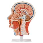
Human Half Head and Neck
Product Code : JL-AM-5983
Life size model showing the outer superficial muscles, vessels and nerves and head neck in one side. View Details
Life size model showing the outer superficial muscles, vessels and nerves and head neck in one side. View Details
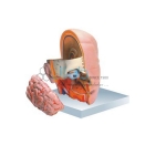
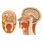
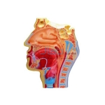
Human Head with Brain
Product Code : JL-AM-5984
Life size 3 part model of the human head which has a removable brain half and the other side exposes the nose, mouth cavity, pharynx, occiput and skull base. View Details
Life size 3 part model of the human head which has a removable brain half and the other side exposes the nose, mouth cavity, pharynx, occiput and skull base. View Details
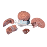
Human Brain Model
Product Code : JL-AM-5986
Life size 4 part model of the human brain. The model is a median section. The right hemisphere has the cerebellum and the stem with occipital lobe. View Details
Life size 4 part model of the human brain. The model is a median section. The right hemisphere has the cerebellum and the stem with occipital lobe. View Details
Human Nose LS
Product Code : JL-AM-5987
Enlarged approx. 2 times, crossing on the windpipe and esophagus can be easily demonstrated. On base. View Details
Enlarged approx. 2 times, crossing on the windpipe and esophagus can be easily demonstrated. On base. View Details
Human Teeth Model
Product Code : JL-AM-5992
2 times enlarged, set of 25 teeth case in break resistant material having accurate anatomical details. View Details
2 times enlarged, set of 25 teeth case in break resistant material having accurate anatomical details. View Details
Human Teeth
Product Code : JL-AM-5993
2 times enlarged, set of 16 teeth case in break resistant material having accurate anatomical details, complete set as in half of upper lower jaw. View Details
2 times enlarged, set of 16 teeth case in break resistant material having accurate anatomical details, complete set as in half of upper lower jaw. View Details

Human Heart
Product Code : JL-AM-5997
3 - times enlarged, dissectable into 2 parts. Showing clearly the chief blood vessels. Structure of auricles and ventricles. View Details
3 - times enlarged, dissectable into 2 parts. Showing clearly the chief blood vessels. Structure of auricles and ventricles. View Details
Human Heart- 4 Parts
Product Code : JL-AM-5999
Enlarged Science Labs, sectioned so that both ventricles and atria open to expose the valves. Large blood vessels near the heart and musculature of the heart are shown. View Details
Enlarged Science Labs, sectioned so that both ventricles and atria open to expose the valves. Large blood vessels near the heart and musculature of the heart are shown. View Details
Human Heart Muscle
Product Code : JL-AM-6001
Model picture showing ultra structure of human heart muscle. Size of the model is 22 x 22 x 37. View Details
Model picture showing ultra structure of human heart muscle. Size of the model is 22 x 22 x 37. View Details
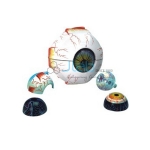
Human Eye 5 Times
Product Code : JL-AM-6002
Enlarged 5 times, 7 parts. After removing the upper half of the sclera (outer shell), the choroids with its verticose veins is exposed, with the removal of second shell (the choroids) details of spot is quite visible. View Details
Enlarged 5 times, 7 parts. After removing the upper half of the sclera (outer shell), the choroids with its verticose veins is exposed, with the removal of second shell (the choroids) details of spot is quite visible. View Details
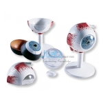
Human Eye Model
Product Code : JL-AM-6003
Human Eye Model-3 Times, full Size -7 Parts The upper half of the sclera with cornea and eye muscle attachments, both halves of choroids with iris and retina, lens and vitreous humor are removable. View Details
Human Eye Model-3 Times, full Size -7 Parts The upper half of the sclera with cornea and eye muscle attachments, both halves of choroids with iris and retina, lens and vitreous humor are removable. View Details
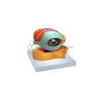
Human Eye with Lid
Product Code : JL-AM-6006
Enlarged 5 times model of the human eye, with 8 parts in bony orbit. Shows eyelid, lachrymal & system and other features around eye ball. View Details
Enlarged 5 times model of the human eye, with 8 parts in bony orbit. Shows eyelid, lachrymal & system and other features around eye ball. View Details
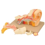
Human Eye Demonstration
Product Code : JL-AM-6007
Science Labs Model Enlarged 5 times, showing the eye socket with the sagittal cutaway. View Details
Science Labs Model Enlarged 5 times, showing the eye socket with the sagittal cutaway. View Details
Human Ear-5 Parts
Product Code : JL-AM-6013
Science Labs Enlarged approximately 4 times. The petros portion of the temporal bone section of the auditory canal are removable, with labyrinth can be taken out and opened. View Details
Science Labs Enlarged approximately 4 times. The petros portion of the temporal bone section of the auditory canal are removable, with labyrinth can be taken out and opened. View Details
Human Ear-6 Parts
Product Code : JL-AM-6015
Science Labs Enlarged 3 times, dissects into 6 parts. Shows the external auditory duct, middle and inner ear with dissectible. View Details
Science Labs Enlarged 3 times, dissects into 6 parts. Shows the external auditory duct, middle and inner ear with dissectible. View Details
Human Digestive Canal
Product Code : JL-AM-6017
Science Labs Showing the structure of Human Digestive Canal, supplied in three parts as illustrated. View Details
Science Labs Showing the structure of Human Digestive Canal, supplied in three parts as illustrated. View Details
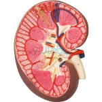
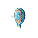
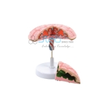
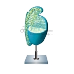
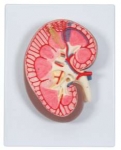
Human Kidney Section
Product Code : JL-AM-6018
Science Labs Longitudinal section of the right kidney. Model shown kidney glomerulus, tubes, one collection tube, pyramids, kidneys orifice system, kidney pelvis, View Details
Science Labs Longitudinal section of the right kidney. Model shown kidney glomerulus, tubes, one collection tube, pyramids, kidneys orifice system, kidney pelvis, View Details
Human Bladder
Product Code : JL-AM-6019
Science Labs Picture shows the seven parts of the human bladder model. Mouted on base. View Details
Science Labs Picture shows the seven parts of the human bladder model. Mouted on base. View Details
Human Abdomen Section
Product Code : JL-AM-6020
Science Labs traverse section model of human abdomen at level of omentum foramen. View Details
Science Labs traverse section model of human abdomen at level of omentum foramen. View Details
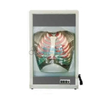
Human Lung Right
Product Code : JL-AM-6021
Science Labs Dissectable into 2 parts showing all the important parts. On base. View Details
Science Labs Dissectable into 2 parts showing all the important parts. On base. View Details
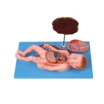
Human Fetus
Product Code : JL-AM-6022
Science Labs Model of human fetus, in 3 parts, shows the relationship among the fetus, fetus membrane and uterus. On Base. View Details
Science Labs Model of human fetus, in 3 parts, shows the relationship among the fetus, fetus membrane and uterus. On Base. View Details
Human Fetal Circulation
Product Code : JL-AM-6023
Science Labs Model of human fetal circulation. On Base. View Details
Science Labs Model of human fetal circulation. On Base. View Details
Human Urinary Organs
Product Code : JL-AM-6026
Science Labs Natural size separates into 3 parts. Kidneys ureters, adrenal glands, bladder with prostate and major blood vessels are shown. View Details
Science Labs Natural size separates into 3 parts. Kidneys ureters, adrenal glands, bladder with prostate and major blood vessels are shown. View Details

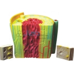
Human Skin
Product Code : JL-AM-6029
Science Labs 75 times life-size, detail structure of 3 layers of the hairly skin in different planes of section are shown. Models show hair follicles with sebaceous, sweat glands. View Details
Science Labs 75 times life-size, detail structure of 3 layers of the hairly skin in different planes of section are shown. Models show hair follicles with sebaceous, sweat glands. View Details
Human Skin-4 Parts
Product Code : JL-AM-6030
Science Labs Enlarged approx 70 times. Layers of the skin can be removed. Showing first rudiments of hair (exposed and in section), sweat gland and sense organs of skin. View Details
Science Labs Enlarged approx 70 times. Layers of the skin can be removed. Showing first rudiments of hair (exposed and in section), sweat gland and sense organs of skin. View Details
Human Brain Stem
Product Code : JL-AM-6042
Science Labs Model of brain stem including transverse sections. View Details
Science Labs Model of brain stem including transverse sections. View Details
Human Long Backbone
Product Code : JL-AM-6043
Science Labs Model of human long backbone and lamina. On base. View Details
Science Labs Model of human long backbone and lamina. On base. View Details
Human Placenta
Product Code : JL-AM-6050
Model of human Placenta and umbilical cord. On base. View Details
Model of human Placenta and umbilical cord. On base. View Details
Human Spinal Cord and Vertebra
Product Code : JL-AM-6051
Expansion Model of transverse section of spinal cord and the vertebra in 2 parts. View Details
Expansion Model of transverse section of spinal cord and the vertebra in 2 parts. View Details
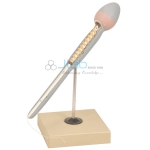
Human Tooth Model
Product Code : JL-AM-6053
Molar tooth dissects into 2 parts, showing the internal structure. On base. Numbered with English Key Card. View Details
Molar tooth dissects into 2 parts, showing the internal structure. On base. Numbered with English Key Card. View Details
Human Foot Bow
Product Code : JL-AM-6054
Model of human foot bow. Set of 3 models, on base. View Details
Model of human foot bow. Set of 3 models, on base. View Details

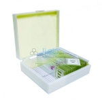
Human Histology Slide Set of 25
Product Code : JL-CE-6722
Set of 25 glass microscope slides showing a varied cross-section of human histology:- View Details
Set of 25 glass microscope slides showing a varied cross-section of human histology:- View Details
Microslide Basic Human Histology
Product Code : JL-CE-6733
Microslides are sets of 8 related 35mm images as photographed through a microscope to be viewed through a Microslide Viewer JLab. View Details
Microslides are sets of 8 related 35mm images as photographed through a microscope to be viewed through a Microslide Viewer JLab. View Details
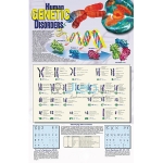
Human Genetic Disorders Poster
Product Code : JL-GAIE-6895
A visual guide to the human genome Detailed view of all 23 human chromosome pairs and the location of the genes which cause some of the most common genetic disorders discovered, to date, as part of the Human Genome Project. View Details
A visual guide to the human genome Detailed view of all 23 human chromosome pairs and the location of the genes which cause some of the most common genetic disorders discovered, to date, as part of the Human Genome Project. View Details
Human Diseases Microslide
Product Code : JL-HE-6930
Bacterial Diseases - Tuberculosis (100x & 1000x), Viral Deseases - Influenza Virus (320,000x), Protist Diseases - Amoebic Dysentery (100x), Fungal Diseases - Trichina Worm (65x) View Details
Bacterial Diseases - Tuberculosis (100x & 1000x), Viral Deseases - Influenza Virus (320,000x), Protist Diseases - Amoebic Dysentery (100x), Fungal Diseases - Trichina Worm (65x) View Details
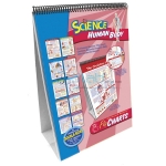
Human Body Flip Chart
Product Code : JL-HBE-6959
Middle School Human Body Flip Chart - Bones, Muscles & Skin; The Circulatory System; The Digestive System; Nutrition; The Immune System; The Digestive System; Nutrition; The Respiratory System View Details
Middle School Human Body Flip Chart - Bones, Muscles & Skin; The Circulatory System; The Digestive System; Nutrition; The Immune System; The Digestive System; Nutrition; The Respiratory System View Details
Human Heart Walk Thru
Product Code : JL-HBE-6961
Book illustrations and hand-out diagrams for the human heart are boring. View Details
Book illustrations and hand-out diagrams for the human heart are boring. View Details
Human Body System Model
Product Code : JL-HBE-6962
Human Body System Model Activity Sets (6 sets) illustrate the functions of the body's major systems. View Details
Human Body System Model Activity Sets (6 sets) illustrate the functions of the body's major systems. View Details
Human Reproduction Microslide
Product Code : JL-HBE-6972
Sperm Cell (1800x) Zygote (200x) Two-celled Embryo - First Cleavage Chromosomes - Squash (400x) View Details
Sperm Cell (1800x) Zygote (200x) Two-celled Embryo - First Cleavage Chromosomes - Squash (400x) View Details
The Digestive System Human Microslide
Product Code : JL-HBE-6982
Microslides are sets of 8 related 35mm images as photographed through a microscope to be viewed through a Microslide Viewer JLab. View Details
Microslides are sets of 8 related 35mm images as photographed through a microscope to be viewed through a Microslide Viewer JLab. View Details
Human Circulatory System Model
Product Code : JL-HBE-7003
Explaining complete circulation in veins and arteries. View Details
Explaining complete circulation in veins and arteries. View Details
Human Digestive System Model
Product Code : JL-HBE-7009
Representation of nose, mouth cavity and oesophagus, pharynx, the gastrointestinal tract, liver with gall bladder, pancreas and spleen. View Details
Representation of nose, mouth cavity and oesophagus, pharynx, the gastrointestinal tract, liver with gall bladder, pancreas and spleen. View Details
Human Ear Model
Product Code : JL-HBE-7012
The model is 2 times the full size and dissects into 3 parts, showing external, middle and internal portions. View Details
The model is 2 times the full size and dissects into 3 parts, showing external, middle and internal portions. View Details

Human Eye Model
Product Code : JL-HBE-7015
Eye Model 6 parts 5 times This model is approximately five times full size. View Details
Eye Model 6 parts 5 times This model is approximately five times full size. View Details
Human Heart Model
Product Code : JL-HBE-7020
Human Heart Model 2 parts Natural Size, front of heart is removable to view the chambers. View Details
Human Heart Model 2 parts Natural Size, front of heart is removable to view the chambers. View Details

Human Elbow Joint Model
Product Code : JL-HBE-7025
A Model of the human elbow joint, showing the essential internal structure, mounted on a base. View Details
A Model of the human elbow joint, showing the essential internal structure, mounted on a base. View Details
Human Foetus Model
Product Code : JL-HBE-7026
JLab three part model of the human foetus showing the relationship between the foetus, foetus membrane and the uterus. View Details
JLab three part model of the human foetus showing the relationship between the foetus, foetus membrane and the uterus. View Details
Human Heart Muscle Model
Product Code : JL-HBE-7027
220 x 220 x 370mm. Shows ultrastructure of the human heart muscle. On base, numbered with key card. View Details
220 x 220 x 370mm. Shows ultrastructure of the human heart muscle. On base, numbered with key card. View Details
Human Knee Joint Model
Product Code : JL-HBE-7029
A model of the Human knee Joint, Showing the essential internal structure, mounted on a base. View Details
A model of the Human knee Joint, Showing the essential internal structure, mounted on a base. View Details
Human Upper and Lower Jaw Model
Product Code : JL-HBE-7033
This model is 3 times full size Showing the tooth roots, spongiosa, vessels and nerves. View Details
This model is 3 times full size Showing the tooth roots, spongiosa, vessels and nerves. View Details
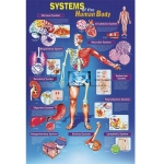
Systems of the Human Body Poster
Product Code : JL-HBE-7089
Dimensions:- Size 890mm W x 585mm H. Unique anatomical perspectives of major human body systems are presented in a clear, concise format for contrast and comparison. View Details
Dimensions:- Size 890mm W x 585mm H. Unique anatomical perspectives of major human body systems are presented in a clear, concise format for contrast and comparison. View Details

Human Diseases Prepared Slide
Product Code : JL-PS-8165
High-quality yet affordable, this prepared slide set is great for teaching students about deadly human diseases. View Details
High-quality yet affordable, this prepared slide set is great for teaching students about deadly human diseases. View Details
Human Pathology Prepared Slide
Product Code : JL-PS-8167
High-quality yet affordable, this prepared slide set is great for introducing human pathology to students. View Details
High-quality yet affordable, this prepared slide set is great for introducing human pathology to students. View Details

Human Physiology Prepared Slide
Product Code : JL-PS-8168
High-quality yet affordable, this human physiology prepared slide set is great for introducing the world of microscopy to students. View Details
High-quality yet affordable, this human physiology prepared slide set is great for introducing the world of microscopy to students. View Details
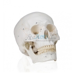
Life Size Human Skull
Product Code : JL-AM-8265
This high-quality human skull model is life-sized and designed with precision to include extremely accurate representations View Details
This high-quality human skull model is life-sized and designed with precision to include extremely accurate representations View Details
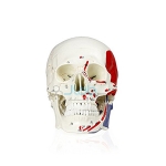
Human Skull with Markings, Muscles and Sutures
Product Code : JL-AM-8266
This 3 part human skull has numerical markings for easy identification of the cranial bones, sutures, and muscles. View Details
This 3 part human skull has numerical markings for easy identification of the cranial bones, sutures, and muscles. View Details
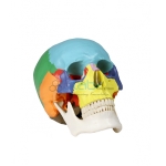
Colored Human Skull
Product Code : JL-AM-8268
This life-size model comes with an instruction booklet that corresponds to the 22 colored sections View Details
This life-size model comes with an instruction booklet that corresponds to the 22 colored sections View Details
Numbered Human Skull
Product Code : JL-AM-8272
Skull sutures, fissures, foramina, and processes are drawn and clearly defined to make the anatomy of the human skull View Details
Skull sutures, fissures, foramina, and processes are drawn and clearly defined to make the anatomy of the human skull View Details
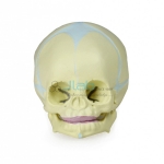
Human Fetal Skull
Product Code : JL-AM-8273
The fontanelle, which becomes bone over time is visible and allows students to study the anatomy of the fetal skull with ease. View Details
The fontanelle, which becomes bone over time is visible and allows students to study the anatomy of the fetal skull with ease. View Details
Human Skull with Brain
Product Code : JL-AM-8275
This human skull model is life-sized and has accurate representations of all sutures, fissures, joints, foramina, and processes. View Details
This human skull model is life-sized and has accurate representations of all sutures, fissures, joints, foramina, and processes. View Details
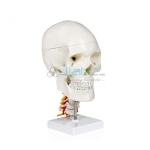
Human Skull with Cervical Spine
Product Code : JL-AM-8276
JLab life-size skull is highly detailed with representations of the fissures, processes, foramina, sutures and cervical spine View Details
JLab life-size skull is highly detailed with representations of the fissures, processes, foramina, sutures and cervical spine View Details
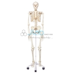
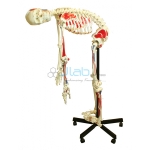
Human Skeleton 66 Full Size 168cm
Product Code : JL-HM-8278
The full-size human skeleton is a great teaching tool in any classroom or laboratory. View Details
The full-size human skeleton is a great teaching tool in any classroom or laboratory. View Details
Human Skeleton Hanging Muscle and Thick Zip Dust Cover
Product Code : JL-HM-8279
The full-size human skeleton is a great teaching tool in any classroom or laboratory. View Details
The full-size human skeleton is a great teaching tool in any classroom or laboratory. View Details
Human Skeleton Full Size with Thick Zip Dust Cover
Product Code : JL-HM-8280
The full-size human skeleton is a great teaching tool in any classroom. View Details
The full-size human skeleton is a great teaching tool in any classroom. View Details
Half Size Human Skeleton 84cm
Product Code : JL-HM-8282
An exceptional price for this 3-foot skeleton mounted on a heavy metal stand. View Details
An exceptional price for this 3-foot skeleton mounted on a heavy metal stand. View Details
Half Size Human Skeleton with Muscle
Product Code : JL-HM-8284
The arms and legs are held on by small screws, making it easy to bend the extremities in all directions. View Details
The arms and legs are held on by small screws, making it easy to bend the extremities in all directions. View Details
Half Size Human Skeleton 84cm with Nerve Ending
Product Code : JL-HM-8285
This affordable teaching skeleton is wired for natural movement and highlights a removable skull with the calvarium cut. View Details
This affordable teaching skeleton is wired for natural movement and highlights a removable skull with the calvarium cut. View Details
Human Skeleton Full Size 170cm Muscles and Ligaments
Product Code : JL-HM-8286
This full-size human skeleton is a great teaching tool in any classroom. View Details
This full-size human skeleton is a great teaching tool in any classroom. View Details
Human Skeleton 67 Full Size Flexible 170cm
Product Code : JL-HM-8287
This full-size human skeleton has a flexible spine that can bend to mimic natural human movement. View Details
This full-size human skeleton has a flexible spine that can bend to mimic natural human movement. View Details
Human Skeleton Full Size Flexible with Muscle 170cm
Product Code : JL-HM-8288
All movable parts are joined by connectors and utilized in such a way to allow movement around the joint. View Details
All movable parts are joined by connectors and utilized in such a way to allow movement around the joint. View Details
Human Spinal Column
Product Code : JL-AM-8290
This spinal column 50cm with occipital bone consists of 24 vertebrae with soft vinyl discs, sacrum, coccyx, and pelvic bones with femur heads. View Details
This spinal column 50cm with occipital bone consists of 24 vertebrae with soft vinyl discs, sacrum, coccyx, and pelvic bones with femur heads. View Details
Human Brain Color Coded 4 Part
Product Code : JL-BMNM-8300
This double life size human brain comes in 4 parts that are color-coded to corresponding sensory regions, functional centers View Details
This double life size human brain comes in 4 parts that are color-coded to corresponding sensory regions, functional centers View Details
Human Respiratory System with Magnified Alveolus
Product Code : JL-AM-8326
The upper respiratory section is medially sectioned to show the interior anatomical details. View Details
The upper respiratory section is medially sectioned to show the interior anatomical details. View Details
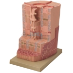
Human Scalp Skin Section Model
Product Code : JL-AM-8345
This relief model displays contrasting colors of the three skin layers-epidermis, derm, and subcutis, with nerves, sweat glands, hair roots, and vessels. View Details
This relief model displays contrasting colors of the three skin layers-epidermis, derm, and subcutis, with nerves, sweat glands, hair roots, and vessels. View Details
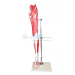
Muscles of Human Leg Model
Product Code : JL-AM-8353
Easily examine the deep leg muscles by removing the superficial muscles. View Details
Easily examine the deep leg muscles by removing the superficial muscles. View Details

Muscles of Human Arm Model
Product Code : JL-AM-8359
The hand and shoulder are also well presented for detailed study of the muscles comprising the rotator cuff and hand. View Details
The hand and shoulder are also well presented for detailed study of the muscles comprising the rotator cuff and hand. View Details
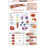
Human Blood Chart
Product Code : JL-ZC-9330
Charts are printed on art paper in full color. Using the latest printing technology. View Details
Charts are printed on art paper in full color. Using the latest printing technology. View Details
Biology Models Mini Human Torso
Product Code : EL-BL-11341
Made of Fibre Glass. Various details shown include: View Details
Made of Fibre Glass. Various details shown include: View Details

Human Excretory System
Product Code : EL-BL-11345
Made of Fibre Glass. Various details shown include: View Details
Made of Fibre Glass. Various details shown include: View Details
Human Urinary System
Product Code : EL-BL-11346
Model is made of advanced HSP resin to maintain long durability. View Details
Model is made of advanced HSP resin to maintain long durability. View Details
Human Kidney
Product Code : EL-BL-11347
Made of Fibre Glass. All-important structures are numbered and identified. Various details shown include: - View Details
Made of Fibre Glass. All-important structures are numbered and identified. Various details shown include: - View Details
Human Uropoiesis
Product Code : EL-BL-11356
Made of Fibre Glass. Uropoiesis is production & excretion of urine from body. Various details shown include: View Details
Made of Fibre Glass. Uropoiesis is production & excretion of urine from body. Various details shown include: View Details
Human Skin
Product Code : EL-BL-11360
Made of Fibre Glass. Various details shown include: View Details
Made of Fibre Glass. Various details shown include: View Details
Human Brain 4 Parts
Product Code : EL-BL-11365
Made of Fiber Glass. All structures are numbered and identified. View Details
Made of Fiber Glass. All structures are numbered and identified. View Details
Mini Human Torso
Product Code : EL-BL-11381
Made of Fiber Glass Various details shown include: View Details
Made of Fiber Glass Various details shown include: View Details
Anatomical Human Skeleton
Product Code : EL-LE-11810
DESCRIPTION :- • It has a movable folding stand. Skeleton is complete up to Phalanges. • Every part is made in a manner so that all joints move freely. View Details
DESCRIPTION :- • It has a movable folding stand. Skeleton is complete up to Phalanges. • Every part is made in a manner so that all joints move freely. View Details
Human Reproductive System Male
Product Code : EL-BL-12066
• Type : Chart • Material : polyart View Details
• Type : Chart • Material : polyart View Details
Human Reproductive System (Female)
Product Code : EL-BL-12067
• Type : Chart • Material : polyart View Details
• Type : Chart • Material : polyart View Details
Human Circulatory System
Product Code : EL-BL-12068
• Type : Chart • Material : polyart View Details
• Type : Chart • Material : polyart View Details
Human Respiratory System
Product Code : EL-BL-12069
• Type : Chart • Material : polyart View Details
• Type : Chart • Material : polyart View Details
Human Heart Model
Product Code : EL-BL-12080
• With dissectible parts • Size : 25 x 23 x 23 cm. View Details
• With dissectible parts • Size : 25 x 23 x 23 cm. View Details
Human Eye
Product Code : EL-BL-12081
The model shows the eyeball with optic nerves and muscles in its natural position in the bony orbit. View Details
The model shows the eyeball with optic nerves and muscles in its natural position in the bony orbit. View Details
Human Brain
Product Code : EL-BL-12085
Model (with dissectible parts) includes 8 parts brain : frontal and parietal lobe, temporal View Details
Model (with dissectible parts) includes 8 parts brain : frontal and parietal lobe, temporal View Details
Human Ear
Product Code : EL-BL-12086
Model (with dissectible parts) dissectible in 6 parts. View Details
Model (with dissectible parts) dissectible in 6 parts. View Details
Human Teeth (Denture)
Product Code : EL-BL-12087
Model (with dissectible parts) 2 times full - size. View Details
Model (with dissectible parts) 2 times full - size. View Details
Human Abdomen/Torso
Product Code : EL-BL-12089
Model (with dissectible parts) size : 33.5” tall (85cm), made of pvc plastic. View Details
Model (with dissectible parts) size : 33.5” tall (85cm), made of pvc plastic. View Details
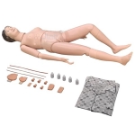
Human Procedure Teaching Model
Product Code : EL-HM-13331
A life-size full body patient care/manikin for nursing procedures. View Details
A life-size full body patient care/manikin for nursing procedures. View Details
Models Human Organ
Product Code : EL-HM-13357
Comprehensive, life-size anatomical teaching model set displaying major human internal organs with detachable and dissectible parts. View Details
Comprehensive, life-size anatomical teaching model set displaying major human internal organs with detachable and dissectible parts. View Details